Registered users receive a variety of benefits including the ability to customize email alerts, create favorite journals list, and save searches.
Please note that a BioOne web account does not automatically grant access to full-text content. An institutional or society member subscription is required to view non-Open Access content.
Contact helpdesk@bioone.org with any questions.
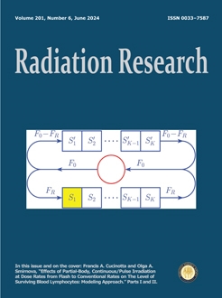
A mathematical model developed by Cucinotta and Smirnova is extended to describe effects of continuous, partial-body irradiation at high doses D and at dose rates N from FLASH to conventional rates on the level of surviving blood lymphocytes in humans and small laboratory animals (mice). Specifically, whereas the applicability of the model is limited to the exposure times shorter than a single cardiac cycle T0, the extended model is capable of describing such effects for the aforementioned and longer exposure times. The extended model is implemented as the algebraic equations. It predicts that the level of surviving blood lymphocytes in humans and mice increases with increasing the dose rate from N = D/T0 to FLASH rates and approaches the upper limiting level of 1 – vR, where vR is the fraction of blood volume in the irradiated part of the blood circulatory system. Levels of surviving blood lymphocytes computed at doses from 10 Gy to 40 Gy and at dose rates N, which equal or exceed 40 Gy/s for humans and 400 Gy/s for mice, are nearly indistinguishable from the upper limiting level. In turn, the level of surviving blood lymphocytes in humans and mice decreases with decreasing the dose rate from N = D/T0 to conventional rates and approaches a lower limiting level. This level strongly depends on the dose D (it is smaller at larger values of D) with a slight dependence on the dose rate N. The model with the parameters specified for mice (together with the model of the dynamics of lymphopoietic system in mice after partial-body irradiation) reproduce, on a quantitative level, the experimental data, according to which the concentration of blood lymphocytes measured in mice in one day after continuous, partial-body irradiation at a high dose and conventional dose rate is smaller at the larger value of vR. Additionally, the model predicts at the same high dose (>10 Gy) a faster restoration of the blood lymphocyte population in humans exposed to continuous, partial-body irradiation at a FLASH dose rate compared to a conventional dose rate.
Mathematical models, which describe effects of partial-body, two- and multiple-pulse irradiation at high total doses D and at average dose rates N from FLASH to conventional rates on the level of surviving blood lymphocytes in humans and mice, have been developed originating in the previously proposed approach. These models predict that levels of surviving blood lymphocytes in humans and mice increase with increasing the dose rate from N = D/TR (TR is the time of the blood flowing into or out of the irradiated segment of the blood circulatory system) to FLASH rates and approach an upper limiting level equal to (1–vR), where vR is the fraction of blood volume in the irradiated segment of the blood circulatory system. Levels of surviving blood lymphocytes computed at total doses D of 10–40 Gy and at average of dose rates N, which are equal to or exceed 40 Gy/s for humans and 400 Gy/s for mice, are nearly indistinguishable from the upper limiting level. These results can be interpreted as the models reproducing the optimal blood lymphocyte sparing in these mammals after such exposures. With decreasing the dose rate from N = D/TR to conventional rates, at multiple-pulse irradiation the levels of surviving blood lymphocytes in humans and mice decrease to lower limiting levels, whereas at two-pulse irradiation they change cyclically and do not fall below their values for the delivery time equal to TR. Additionally, effects of two- and multiple-pulse irradiation of the whole abdomen in mice on the level of surviving blood lymphocytes are simulated within the developed models. Regimens of two- and multiple-pulse irradiation are taken the same as those reported in experiments, where effects of such exposures on the level of surviving crypts in mice were studied. Juxtaposing the modeling results with the experimental data reveals that the level of surviving blood lymphocytes in mice after two- and multiple-pulse irradiation of the abdomen at average dose rates N from FLASH to conventional rates modulates the level of surviving crypts in these animals after such exposures. A hypothesis is proposed to explain this phenomenon.
There is a need for point-of-care diagnostics for future mass casualty events involving radiation exposure. The development of radiation exposure and dose prediction algorithms for biodosimetry is needed for screening of large populations during these scenarios, and exploration of the potential effects which sex, age, genetic heterogeneity, and physiological comorbidities may have on the utility of biodosimetry diagnostics is needed. In the current study, proteomic profiling was used to examine sex-specific differences in age-matched C57BL6 mice on the blood proteome after radiation exposure, and the usefulness of development and application of biodosimetry algorithms using both male and female samples. Male and female mice between 9–11 weeks of age received a dose of total-body irradiation (TBI) of either 2, 4 or 8 Gy and plasma was collected at days 1, 3 and 7 postirradiation. Plasma was then screened using the SomaScan v4.1 assay for ∼7,000 protein analytes. A subset panel of protein biomarkers demonstrated significant (FDR < 0.05 and |logFC| > 0.2) changes in expression after radiation exposure. All proteins were used for feature selection to build predictive models of radiation exposure using different sample and sex-specific cohorts. Both binary (prediction of any radiation exposure) and multidose (prediction of specific radiation dose) model series were developed using either female and male samples combined or only female or only male samples. The binary series (models 1, 2 and 3) and multidose series (models 4, 5 and 6) included female/male combined, female only and male only respectively. Detectable values were obtained for all ∼7,000 proteins included in the SomaScan assay for all samples. Each model algorithm built using a unique sample cohort was validated with a training set of samples and tested with a separate new sample series. Overall predictive accuracies in the binary model series was ∼100% at the model training level, and when tested with fresh samples, 97.9% for model 1 (female and male) and 100% for model 2 (female only) and model 3 (male only). When sex-specific models 2 and 3 were tested with the opposite sex, the overall predictive accuracy rate dropped to 62.5% for model 2 and remained 100% for model 3. The overall predictive accuracy rate in the multidose model series was 100% for all models at the model training level and, when tested with fresh samples, 83.3%, 75% and 83.3% for Multidose models 4–6, respectively. When sex-specific model 5 (female only) and model 6 (male only) were tested with the opposite sex, the overall predictive accuracy rate dropped to 52.1% and 68.8%, respectively. These models represent novel predictive panels of radiation-responsive proteomic biomarkers and illustrate the utility and necessity of considering sex-specific differences in development of radiation biodosimetry prediction algorithms. As sex-specific differences were observed in this study, and as use of point-of-care radiation diagnostics in future mass casualty settings will necessarily include persons of both sexes, consideration of sex-specific variation is essential to ensure these diagnostic tools have practical utility in the field.
Micronuclei, detected through the cytokinesis-block micronucleus assay, are valuable indicators of ionizing radiation exposure, especially in short-term lymphocyte cultures. The peripheral human blood lymphocyte assay is recognized as a prime candidate for automated biodosimetry. In a prior project at the Columbia University Center for Radiological Research, we automated this assay using the 96-well ANSI/SLAS microplate standard format and relied on established biotech robotic systems named Rapid Automated Biodosimetry Tool (RABiT). In this study, we present the application of a similar automated biotech setup at an external high-throughput facility (RABiT-III) to implement the same automated cytokinesis-block micronucleus assay. Specifically, we employed the Agilent BRAVO liquid-handling system and GE IN Cell Analyzer 6000 imaging system in conjunction with the PerkinElmer Columbus image data storage and analysis system. Notably, this analysis system features an embedded PhenoLOGIC machine learning module, simplifying the creation of cell classification algorithms for CBMN assay image analysis and enabling the generation of radiation dose-response curves. This investigation underscores the adaptability of the RABiT-II CBMN protocol to diverse RABiT-III biotech robotic platforms in non-specialized biodosimetry centers. Furthermore, it highlights the advantages of machine learning in rapidly developing algorithms crucial for the high-throughput automated analysis of RABiT-III images.
Radiation enteritis is a common complication of abdominal and pelvic radiotherapy. Several previous studies showed that fecal microbiota transplantation (FMT) could alleviate radiation enteritis. In this study, we investigated the efficacy of FMT in alleviating radiation enteritis and explored the mechanisms by multi-omics approaches. Briefly, C57BL/6J mice were subjected to 9 Gy irradiation to the localized abdominal field, and randomized received FMT from healthy donor mice or saline. H&E staining of harvested small intestine showed FMT decreased epithelial injury. Radiation-induced microbiota dysbiosis, characterized by a decrease in beneficial bacteria Lactobacillaceae and Lachnospiraceae, while these bacteria were restored by FMT. Fecal metabolomics analysis revealed that FMT modulated metabolic dysregulation. Two tryptophan pathway metabolites, indole-3-acetaldehyde and N-Acetyl-5-hydroxytryptamine were decreased after irradiation, whereas these metabolites showed a pronounced recovery in mice receiving FMT. Proteomics analysis of small intestine indicated that radiation enteritis triggered immune-inflammatory responses, which were potentially mitigated by FMT. In 21 patients receiving pelvic radiotherapy for cervical cancer, those who developed enteritis (n = 15) had higher abundance in Lachnospiraceae. Moreover, Indole-3-acetaldehyde was reduced after irradiation. These findings provide insights into the therapeutic effects of FMT in radiation enteritis and highlight Lachnospiraceae and the tryptophan metabolite, Indole-3-acetaldehyde may protect against radiation enteritis.
The linear, non-threshold (LNT) hypothesis of cancer induction derived from studies of populations exposed to moderate-to-high acute radiation doses may not be indicative of cancer risks associated with lifetime radiation exposures less than 100 mSv. The objective of this study was to examine risks and dose-response patterns of lymphohematopoietic cancer (LHC) and its types associated with low radiation exposure while adjusting for possible confounding factors. A retrospective cohort of 437,937 U.S. nuclear shipyard workers (153,930 radiation and 284,007 non-radiation workers) was followed from 1957 to 2011, with 3,699 LHC deaths observed. The risk of LHC in radiation workers was initially compared to the risk in non-radiation workers. Time dependent accumulated radiation dose, lagged 2 years, was used in categorical and continuous dose analysis among radiation workers to examine the LHC risks and possible dose-response relationships based on Poisson regression models. These analyses controlled for sex, race, time dependent age, calendar time, socioeconomic status, solvent-related last job, and age at first hire. The median lifetime radiation dose for the radiation worker population was 0.82 mSv and the 95th percentile dose was 83.63 mSv. The study shows: 1. LHC mortality for radiation workers was significantly lower than non-radiation workers relative risk: 0.927; 95% confidence intervals (95% CI): 0.865, 0.992; P = 0.030]. Among LHC types, the risks for lymphoid leukemia and lymphomas in radiation workers were lower than the risk in non-radiation workers with statistical significance, while the risk for the rest of LHC types did not show any statistically significant difference. 2. In categorical dose analysis among radiation workers, sample size weighted linear trend of relative risk (RRs) for LHC and its types in five dose categories (>0–<25, 25–<50, 50–<100, 100–<200, and > = 200 mSv) vs. 0 mSv were not statistically significant, although there was an elevation of RR for chronic myeloid leukemia only in the 50-<100 mSv category (RR: 2.746; 95% CI: 1.002, 7.521; P = 0.049) vs. 0 mSv. 3. The Poisson regression analyses among radiation workers using the time dependent radiation dose as a continuous variable showed an excess relative risk (ERR) for LHC at 100 mSv of 0.094 (95% CI: –0.037, 0.225; P = 0.158) and leukemia less chronic lymphoid leukemia, of 0.178 (95% CI: –0.085, 0.440; P = 0.440) vs. 0 mSv. The ERRs and their linear trend for all other types were not statistically significant.
This study offers a review of published data on DNA double strand break (DSB) repair kinetics after exposure to ionizing radiation. By compiling a database, which currently includes 285 DNA DSB repair experiments utilizing both photons and ions, we investigate the impact of distinct experimental parameters on the kinetics of DNA DSB repair. Methodological differences and inconsistencies in reporting make the comparison of data generated by different research groups challenging. Nevertheless, by implementing filtering criteria, we can compare repair kinetics obtained with normal and tumor cells derived from human or animal tissues, as well as cells exposed to photons or ions ranging from hydrogen to iron ions. In addition, several repair curves of repair deficient cell lines were included. The study aims to provide researchers with a comprehensive overview of experimental factors that may confound results and emphasize the importance of precise reporting of experimental parameters. Moreover, we identify gaps in the literature that require attention in future studies, aiming to address clinically relevant questions related to radiotherapy. The database can be freely accessed at: https://github.com/weradstake/DRDNA.
This review focuses on early discoveries that contributed to our understanding and the scope of transcriptional responses after radiation damage. Before the development of modern approaches to assess overall global transcriptomic responses, the idea that mammalian cells could respond to DNA-damaging agents in a manner analogous to bacteria was not generally accepted. To investigate this possibility, the development of technology to identify differentially expressed low-abundance transcripts substantially facilitated our appreciation that DNA damaging agents like UV radiation and subsequently ionizing radiation did in fact produce robust transcriptional responses. Here we focus on our identification and characterization of radiation-inducible genes, and how even early studies on stress gene signaling highlighted the broad scope of transcriptional responses to radiation damage. Since then, the central role of transcriptional responses to radiation injury in maintaining genome integrity has been highlighted in many processes, including cell cycle checkpoint control, resistance to cancer by p53 and other key factors, cell senescence, and metabolism.
Thomas A. Winters, Libero Marzella, Olivia Molinar-Inglis, Paul W. Price, Nyun Calvin Han, Jonathan E. Cohen, Sue-Jane Wang, Anthony F. Fotenos, Julie M. Sullivan, John I. Esker, Paula J. Lapinskas, Andrea L. DiCarlo
There have been a number of reported human exposures to high dose radiation, resulting from accidents at nuclear power plants (e.g., Chernobyl), atomic bombings (Hiroshima and Nagasaki), and mishaps in industrial and medical settings. If absorbed radiation doses are high enough, evolution of acute radiation syndromes (ARS) will likely impact both the bone marrow as well as the gastrointestinal (GI) tract. Damage incurred in the latter can lead to nutrient malabsorption, dehydration, electrolyte imbalance, altered microbiome and metabolites, and impaired barrier function, which can lead to septicemia and death. To prepare for a medical response should such an incident arise, the National Institute of Allergy and Infectious Diseases (NIAID) funds basic and translational research to address radiation-induced GI-ARS, which remains a critical and prioritized unmet need. Areas of interest include identification of targets for damage and mitigation, animal model development, and testing of medical countermeasures (MCMs) to address GI complications resulting from radiation exposure. To appropriately model expected human responses, it is helpful to study analogous disease states in the clinic that resemble GI-ARS, to inform on best practices for diagnosis and treatment, and translate them back to inform nonclinical drug efficacy models. For these reasons, the NIAID partnered with two other U.S. government agencies (the Biomedical Advanced Research and Development Authority, and the Food and Drug Administration), to explore models, biomarkers, and diagnostics to improve understanding of the complexities of GI-ARS and investigate promising treatment approaches. A two-day workshop was convened in August 2022 that comprised presentations from academia, industry, healthcare, and government, and highlighted talks from 26 subject matter experts across five scientific sessions. This report provides an overview of information that was presented during the conference, and important discussions surrounding a broad range of topics that are critical for the research, development, licensure, and use of MCMs for GI-ARS.
This article is only available to subscribers. It is not available for individual sale.
Access to the requested content is limited to institutions that have
purchased or subscribe to this BioOne eBook Collection. You are receiving
this notice because your organization may not have this eBook access.*
*Shibboleth/Open Athens users-please
sign in
to access your institution's subscriptions.
Additional information about institution subscriptions can be foundhere