Registered users receive a variety of benefits including the ability to customize email alerts, create favorite journals list, and save searches.
Please note that a BioOne web account does not automatically grant access to full-text content. An institutional or society member subscription is required to view non-Open Access content.
Contact helpdesk@bioone.org with any questions.
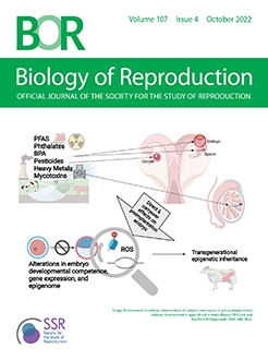
Environmental impacts on reproductive function are well documented in humans, yet little information is known about the effects on large animals. The interface of environment and reproduction has evolved prudently with a concerted effort to ensure global food sustainability tightly integrated with the application of technological advances in agriculture production that include nutrient and resource management. Exposure to environmental toxicants through chemical pesticide application and industry practices has coincided with a decline in cattle and human fertility. The increased adoption of agriculture animals for human biomedical models further emphasizes the importance of understanding the consequences of livestock exposure to environmentally and physiologically relevant levels of contaminants to preimplantation embryo development. In addition, increased awareness of paternal contributions to the early embryo that include both genetic and nongenetic factors supports the need to define environmental interactions from gamete to genome. Herein we summarize current knowledge of common environmental contaminants on reproductive function including direct and indirect effects on embryo development success in livestock. Information obtained from a diverse number of species including humans is presented to illustrate gaps in knowledge within livestock directly pertaining to agriculture success, sustainability, clinical practice, and biomedical research.
Summary Sentence
Livestock embryo exposure to environmental contaminants suggests detrimental effects to developmental competence that remain largely uncharacterized.
Endometriosis that afflicts one in 10 women of reproductive age is characterized by growth of endometrial tissue in the extra-uterine sites and encompasses metabolic-, immunologic-, and endocrine-disruption. Importantly, several comorbidities are associated with endometriosis, especially autoimmune disorders such as inflammatory bowel disease. Primarily thought of as a condition arising from retrograde menstruation, emerging evidence uncovered a functional link between the gut microbiota and endometriosis. Specifically, recent findings revealed altered gut microbiota profiles in endometriosis and in turn this altered microbiota appears to be causal in the disease progression, implying a bidirectional crosstalk. In this review, we discuss the complex etiology and pathogenesis of endometriosis, emphasizing on this recently recognized role of gut microbiome. We review the gut microbiome structure and functions and its complex network of interactions with the host for maintenance of homeostasis that is crucial for disease prevention. We highlight the underlying mechanisms on how some bacteria promote disease progression and others protect against endometriosis. Furthermore, we highlight the areas that require future emphases in the gut microbiome–endometriosis nexus and the potential microbiome-based therapies for amelioration of endometriosis.
Summary Sentence
The commensal gut microbes prevent endometriotic lesions formation through protective effect of short chain fatty acids (SCFAs) while other gut microbes may promote lesion formation in the state of dysbiosis through disruption of gut barrier integrity and the resulting macrophage activation.
Phosphoinositides (PIs) are relatively rare lipid components of the cellular membranes. Their homeostasis is tightly controlled by specific PI kinases and PI phosphatases. PIs play essential roles in cellular signaling, cytoskeletal organization, and secretory processes in various diseases and normal physiology. Gene targeting experiments strongly suggest that in mice with deficiency of several PI phosphatases, such as Pten, Mtmrs, Inpp4b, and Inpp5b, spermatogenesis is affected, resulting in partial or complete infertility. Similarly, in men, loss of several of the PI phosphatases is observed in infertility characterized by the lack of mature sperm. Using available gene expression databases, we compare the expression of known PI phosphatases in various testicular cell types, infertility patients, and mouse age-dependent testicular gene expression, and discuss their potential roles in testis physiology and spermatogenesis.
Summary Sentence
Phosphatidylinositol polyphosphate phosphatases (PI phophatases) play an important role in testicular function by regulating spermatogenesis, maintaining testicular muscle function and blood–testis barrier, and by modulating overall metabolic health.
Free amino acids are present in the natural environment of the preimplantation embryo, and their availability can influence early embryo development. Glutamic acid is one of the amino acids with the highest concentrations in female reproductive fluids, and we investigated whether glutamic acid/glutamate can affect preimplantation embryo development by acting through cell membrane receptors. Using reverse transcription-polymerase chain reaction, we detected 15 ionotropic glutamate receptor transcripts and 8 metabotropic glutamate receptor transcripts in mouse ovulated oocytes and/or in vivo developed blastocysts. Using immunohistochemistry, we detected the expression of two α-amino-3-hydroxy-5-methyl-4-isoxazolepropionic acid (AMPA) receptor subunits, three kainate receptor subunits, and member 5 metabotropic glutamate receptor protein in blastocysts. Extracellular concentrations of glutamic acid starting at 5 mM impaired mouse blastocyst development, and this fact may be of great practical importance since glutamic acid and its salts (mainly monosodium glutamate) are widely used as food additives. Experiments with glutamate receptor agonists (in combination with gene expression analysis) revealed that specific AMPA receptors (formed from glutamate receptor, ionotropic, AMPA3 [GRIA3] and/or glutamate receptor, ionotropic, AMPA4 [GRIA4] subunits), kainate receptors (formed from glutamate receptor, ionotropic, kainate 3 [GRIK3] and glutamate receptor, ionotropic, kainate 4 [GRIK4] or glutamate receptor, ionotropic, kainate 5 [GRIK5] subunits), and member 5 metabotropic glutamate receptor (GRM5) were involved in this effect. The glutamic acid-induced effects were prevented or reduced by pretreatment of blastocysts with AMPA, kainate, and GRM5 receptor antagonists, further confirming the involvement of these receptor types. Our results show that glutamic acid can act as a signaling molecule in preimplantation embryos, exerting its effects through the activation of cell membrane receptors.
Summary Sentence
Several types of glutamate receptors are expressed in mouse oocytes and blastocysts, and extracellular concentrations of glutamic acid at 5 mM can inhibit blastocyst development by activating glutamate receptors.
WNT signaling is important for regulation of embryonic development. The most abundant WNT gene expressed in the bovine endometrium during the preimplantation period is WNT5A. One objective was to determine whether WNT5A regulates competence of the bovine preimplantation embryo to become a blastocyst and alters the number of cells in the inner cell mass and trophectoderm. A second objective was to delineate features of the cell-signaling mechanisms involved in WNT5A actions. WNT5A caused a concentration-dependent increase in the proportion of embryos developing to the blastocyst stage and in the number of inner cell mass cells in the resultant blastocysts. A concentration of 200 ng/mL was most effective, and a higher concentration of 400 ng/mL was not stimulatory. Bovine serum albumin in culture reduced the magnitude of effects of WNT5A on development to the blastocyst stage. WNT5A affected expression of 173 genes at the morula stage; all were upregulated by WNT5A. Many of the upregulated genes were associated with cell signaling. Actions of WNT5A on development to the blastocyst stage were suppressed by a Rho-associated coiled-coil kinase (ROCK) signaling inhibitor, suggesting that WNT5A acts through Ras homology gene family member A (RhoA)/ROCK signaling. Other experiments indicated that actions of WNT5A are independent of the canonical β-catenin signaling pathway and RAC1/c-Jun N-terminal kinase (JNK) signaling. This is the first report outlining the actions of WNT5A to alter the development of the mammalian embryo. These findings provide insights into how embryokines regulate maternal–embryonic communication.
Summary Sentence:
WNT5A, one of the most abundant cell-signaling ligands expressed in the endometrium, is an important regulator of early development because it can act on the preimplantation bovine embryo to improve competence to develop to the blastocyst stage and increase the number of cells in inner cell mass.
One mechanism by which the maternal environment regulates the early embryo is by secretion of cell-signaling molecules. One of these is dickkopf WNT signaling pathway inhibitor 1. Objectives were to (A) resolve discrepancies in the literature regarding effects of dickkopf WNT signaling pathway inhibitor 1 in the bovine embryo on development of trophectoderm and competence to establish pregnancy after embryo transfer and (B) determine whether there are long-term consequences of dickkopf WNT signaling pathway inhibitor 1 on placental function and postnatal phenotype. Embryos produced in vitro were cultured with vehicle or 100 ng/mL recombinant human dickkopf WNT signaling pathway inhibitor 1 from Days 5 to 7.5 of development (i.e., the morula and blastocyst stages of development). dickkopf WNT signaling pathway inhibitor 1 increased the number of cells positive for the trophectoderm marker CDX2 at Day 7.5 of development while having no effect on number of cells positive for the inner cell mass marker SOX2. There was no effect of dickkopf WNT signaling pathway inhibitor 1 on pregnancy or calving rate after transfer of blastocysts produced with Y-sorted semen to either lactating dairy cows or suckling beef cows. Treatment with dickkopf WNT signaling pathway inhibitor 1 at the morula-to-blastocyst stages programmed placental function, as measured by an effect of dickkopf WNT signaling pathway inhibitor 1 on plasma concentrations of pregnancy associated glycoproteins and placental lactogen at Day 160 of gestation (although not on other days examined). dickkopf WNT signaling pathway inhibitor 1 treatment also resulted in calves that were heavier at birth as compared to calves derived from control embryos. After birth, dickkopf WNT signaling pathway inhibitor 1 calves grew slower than controls. Results confirm that dickkopf WNT signaling pathway inhibitor 1 alters the developmental program of the bovine embryo to affect both prenatal and postnatal phenotypes.
Summary Sentence
DKK1 can act to alter the early developmental program of the bovine embryo to affect prenatal and postnatal phenotypes.
The processes underlying adenomyosis are similar to those of tumor metastasis, and it is defined as progressive invasion by the endometrium and the subsequent creation of ectopic lesions. GRIM-19 regulates cell death via the mitochondrial respiratory chain. Stress following oxygen deprivation can induce tumor cell autophagy, leading to cell invasion and migration. Here, we revealed that GRIM-19 negatively regulates autophagy, and, at least in adenomyosis, decreased expression of GRIM-19 is accompanied by an increased level of autophagy and 5′-adenosine monophosphate-activated protein kinase-Unc-51 like autophagy activating kinase 1 (AMPK-ULK1) activation. Upregulation of GRIM-19 expression in human primary endometrial cells and ISHIKAWA cells inhibits autophagy via the AMPK-ULK1 pathway and helps control cell invasion and migration. In addition, we also identified increased expression of AMPK and ULK1, and higher levels of autophagy in the uterine tissues of GRIM-19+/– mice. Importantly, the function of the GRIM-19-AMPK-ULK1 axis in regulating autophagy in adenomyosis is similar to that of tumor tissues, which may help elucidate the regulation of adenomyosis tumor-like behavior, and is expected to help identify novel targets for the diagnosis and treatment of adenomyosis.
Summary Sentence
GRIM-19 knockdown increased autophagy and AMPK/ULK expression, whereas GRIM-19 overexpression reduced their expression in vitro; these observations were validated in a GRIM-19 allelic gene heterozygotes mouse model.
Worldwide, cervical artificial insemination using frozen–thawed semen yields low pregnancy rates. The only exception to this is in Norway, where vaginal insemination with frozen–thawed semen yields pregnancy rates in excess of 60% and which has been attributed to the specific ewe breed used. Our previous work demonstrated differences in cervical gene expression at the follicular phase of the estrous cycle in ewe breeds with known differences in pregnancy rates. In this study, we characterized the cervical transcriptome of the same ewe breeds [Suffolk, Belclare, Fur, and Norwegian White Sheep (NWS)] during the luteal phase, as an optimal environment at the luteal phase could better prepare the cervix for sperm migration through the cervix at the subsequent follicular phase. High-quality RNA extracted from postmortem cervical tissue was analyzed by RNA sequencing. After stringent filtering, 1051, 1924, and 611 differentially expressed genes (DEGs) were detected in the low-fertility Suffolk breed compared with Belclare, Fur, and NWS, respectively. Gene ontology analysis identified increased humoral adaptive immune response pathways in Suffolk. Increased expression of multiple immune genes supports the presence of an active immune response in the cervix of Suffolk ewes, which differentiates them significantly from the other three ewe breeds. Inflammatory pathways were upregulated in the Suffolk, resulting in higher expression of the potent pro-inflammatory cytokines. Therefore, higher levels of pro-inflammatory cytokines indicate unresolved inflammation in the cervix of the low-fertility Suffolk breed that could contribute to reduced cervical sperm transport in the next follicular phase.
Summary Sentence
Increased humoral adaptive immune response in the low-fertility Suffolk breed at the luteal phase indicates unresolved inflammation in the cervix of Suffolk that may contribute to reduced cervical sperm transport in the subsequent follicular phase.
Objectives were to test the hypothesis that pre- and post-natal nutrition in the bovine female, independently or interactively, affect age at puberty and functional characteristics of the estrous cycle of sexually mature offspring. Brangus and Braford (n = 97) beef cows bearing a female fetus were fed to achieve body condition scores of 7.5–8 (H, obese), 5.5–6 (M, moderate), or 3–3.5 (L, thin) by the start of the third trimester and maintained until parturition. Heifer offspring were weaned and fed to gain weight at either a high (H; 1 kg/day) or a low (L; 0.5 kg/day) rate between 4 and 8 months of age, then fed the same diet during a common feeding period until puberty, which resulted in compensatory growth of heifers in the L group. Heifers (n = 95) from the H postnatal diet reached puberty 2 months earlier (12 ± 0.4 months; P = 0.0002) than those from the L postnatal diet (14 ± 0.4 months). Estrous cycles of a subgroup of postpubertal heifers (n = 53) were synchronized to evaluate antral follicle count (AFC), rate of growth and size of the pre-ovulatory follicle, size of corpus luteum and ovary, endometrial thickness, and plasma concentrations of progesterone and estradiol-17β (E2). Although there was a trend for postnatal H heifers to have greater AFC and plasma concentrations of E2 compared to L heifers, neither pre- nor post-natal nutrition affected any other physiological or hormonal variables, including short-term fertility. Postnatal nutritional effects on pubertal age remained the dominant observed feature.
Summary Sentence
Age at puberty was advanced in heifers exposed to a high rate of body weight gain during the juvenile period, however neither pre- nor post-natal nutrition affected any other physiological or hormonal variables, including short-term fertility.
The Notch signaling pathway is required for reproductive success. This pathway activates its transcriptional effector, recombination signal binding protein for immunoglobulin kappa J (Rbpj), to induce transcription of its target genes. This signaling pathway is required for successful decidualization, implantation, and uterine repair following parturition. To identify the compartmental specific roles of the Notch signaling pathway in the establishment of pregnancy, we generated epithelial and decidual stromal cell specific knockouts of Rbpj utilizing lactoferrin iCre and Prl8A2 iCre, respectively. Both conditional knockout mouse models were fertile. The Rbpj epithelial knockout mice displayed 27% resorption sites at E15.5, but this did not significantly impact the number of live born pups compared with controls. In addition, the Rbpj epithelial knockout mice displayed increased estrogen signaling in their stromal compartment. Given that both mouse models exhibited fertility comparable to control animals, the epithelial and stromal specific nature of the iCre recombinases utilized, and previously published Rbpj total uterine knockout mouse models, we conclude that Notch effector Rbpj signaling is required at the initiation of pregnancy to support decidualization in stromal cells, but that Rbpj is not required in the epithelial compartment nor is it required for post-implantation pregnancy success.
Summary Sentence
Notch effector Rbpj signaling is required at the initiation of pregnancy to support decidualization in stromal cells, but Rbpj is not required in the epithelial compartment nor is it required for post-implantation pregnancy success.
The composition of cell contacts in the endometrium plays an important role in the process of embryo implantation and the establishment of pregnancy. In previous studies, we showed an induction of the tight junction protein claudin-3 in the developing decidua from day 6.5 of pregnancy onward. To evaluate the role of this specific claudin-3 distribution, we here evaluated the effect of an endometrial claudin-3 deletion in implantation and embryo development in claudin-3 knockout mice. Claudin-3 knockout mice were fertile but revealed a slightly reduced amount of implantation sites as well as of litter size. Though implantation sites showed morphologically regularly developed embryos and deciduas, depth of ectoplacental cone invasion was reduced in tendency compared to controls. The weight of the implantation sites on day 6.5 and 8.5 of pregnancy as well as the weight of the embryos on day 17.5 of pregnancy, but not of the placentas, was significantly reduced in claudin-3 knockout mice due to a maternal effect. This could be due to an impairment of decidualization as substantiated by a downregulation of the transcription of various decidua-associated genes in the early implantation sites of claudin-3 knockout mice. The fact that claudin-3 knockout mice are nevertheless fertile possibly may be compensated by the presence of other claudins like claudin-4 and claudin-10.
Summary Sentence
Though claudin-3-deficient mice are fertile, endometrial deletion of claudin-3 impairs decidualization leading to reduced size of implantation sites in early pregnancy and to a decrease in prenatal fetal weight.
Anamaria-Cristina Herta, Lucia von Mengden, Nazli Akin, Katy Billooye, Wim Coucke, Julia van Leersum, Berta Cava-Cami, Laura Saucedo-Cuevas, Fábio Klamt, Johan Smitz, Ellen Anckaert
Establishing an ideal human follicle culture system for oncofertility patients relies mainly on animal models since donor tissue is scarce and often of suboptimal quality. The in vitro system developed in our laboratory supports the growth of prepubertal mouse secondary follicles up to mature oocytes. Given the importance of glucose in preparing the oocyte for proper maturation, a baseline characterization of follicle metabolism both in the culture system and in vivo was carried out. Markers of glucose-related pathways (glycolysis, tricarboxylic acid [TCA] cycle, pentose phosphate pathway [PPP], polyol pathway, and hexosamine biosynthetic pathway), as well as the antioxidant capacity, were measured in the different follicle cell types by both enzymatic activities (spectrophotometric detection) and gene expression (qPCR). This study confirmed that in vivo the somatic cells, mainly granulosa, exhibit intense glycolytic activity, while oocytes perform PPP. Throughout the final maturation step, oocytes in vivo and in vitro showed steady levels for all the key enzymes and metabolites. On the other hand, ovulation triggers a boost of pyruvate and lactate uptake in cumulus cells in vivo, consumes reduced nicotinamide adenine dinucleotide phosphate, and increases TCA cycle and small molecules antioxidant capacity activities, while in vitro, the metabolic upregulation in all the studied pathways is limited. This altered metabolic pattern might be a consequence of cell exhaustion because of culture conditions, impeding cumulus cells to fulfill their role in providing proper support for acquiring oocyte competence.
Summary Sentence
In vitro-cultured mouse follicles exhibit altered glycolytic activity and redox metabolism in the somatic compartment during meiotic maturation.
Oocyte developmental potential is intimately linked to metabolism. Existing approaches to measure metabolism in the cumulus oocyte complex (COC) do not provide information on the separate cumulus and oocyte compartments. Development of an assay that achieves this may lead to an accurate diagnostic for oocyte quality. Optical imaging of the autofluorescent cofactors reduced nicotinamide adenine dinucleotide (phosphate) [NAD(P)H] and flavin adenine dinucleotide (FAD) provides a spatially resolved indicator of metabolism via the optical redox ratio (FAD/[NAD(P)H + FAD]). This may provide an assessment of oocyte quality. Here, we determined whether the optical redox ratio is a robust methodology for measuring metabolism in the cumulus and oocyte compartments compared with oxygen consumption in the whole COC. We also determined whether optical imaging could detect metabolic differences associated with poor oocyte quality (etomoxir-treated). We used confocal microscopy to measure NAD(P)H and FAD, and extracellular flux to measure oxygen consumption. The optical redox ratio accurately reflected metabolism in the oocyte compartment when compared with oxygen consumption (whole COC). Etomoxir-treated COCs showed significantly lower levels of NAD(P)H and FAD compared to control. We further validated this approach using hyperspectral imaging, which is clinically compatible due to its low energy dose. This confirmed lower NAD(P)H and FAD in etomoxir-treated COCs. When comparing hyperspectral imaged vs non-imaged COCs, subsequent preimplantation development and post-transfer viability were comparable. Collectively, these results demonstrate that label-free optical imaging of metabolic cofactors is a safe and sensitive assay for measuring metabolism and has potential to assess oocyte developmental competence.
Summary Sentence
Optical imaging using confocal and hyperspectral microscopy is able to measure dynamic changes in oocyte metabolism.
G protein-coupled estrogen receptor (GPER), a seven-transmembrane G protein-coupled receptor, mediates the rapid pre-genomic signaling actions of estrogen and derivatives thereof. The expression of GPER is extensive in mammal male reproductive system. However, the functional role of GPER in mouse sperm has not yet been well recognized. This study revealed that GPER was expressed at the acrosome and the mid-flagellum of the mouse sperm. The endogenous GPER ligand 17β-estradiol and the selective GPER agonist G1 increased intracellular Ca2+ concentration ([Ca2+]i) in mouse sperm, which could be abolished by G15, an antagonist of GPER. In addition, the G1-stimulated Ca2+ response was attenuated by interference with the phospholipase C (PLC) signaling pathways or by blocking the cation channel of sperm (CatSper). Chlortetracycline staining assay showed that the activation of GPER increased the incidence of acrosome-reacted sperm. Conclusively, GPER was located at the acrosome and mid-flagellum of the mouse sperm. Activation of GPER triggered the elevation of [Ca2+]i through PLC-dependent Ca2+ mobilization and CatSper-mediated Ca2+ influx, which promoted the acrosome reaction of mouse sperm.
Polycystic ovary syndrome (PCOS) is a common endocrine and metabolic disease in women, with clinical manifestations of anovulation and hyperandrogenaemia. The treatment of PCOS mainly focuses on improving clinical symptoms, such as insulin sensitivity or menstrual disorder, through drug treatment. However, due to the pathogenesis diversity of PCOS, there is still a lack of effective treatment in clinics. Metabolic disorder is the key factor in the occurrence of PCOS. Brown adipose tissue (BAT) is a special adipose tissue in the human body that can participate in metabolic balance by improving heat production. BAT has been demonstrated to be an important substance involved in the metabolic disorder of PCOS. Although increasing evidence indicates that BAT transplantation can improve the symptoms of PCOS, it is difficult to achieve BAT transplantation at present due to technical limitations. Stimulation of BAT activation by exogenous substances may be an effective alternative therapy for PCOS. In this study, we investigated the effects of Irisin on dehydroepiandrosterone (DHEA)-induced PCOS in mice and evaluated the effect of Irisin on serum hormone levels and changes in body temperature, body weight, and ovarian morphology. In our study, we found that Irisin can enhance the thermogenesis and insulin sensitivity of PCOS mice by activating the function of BAT. In addition, Irisin treatment can correct the menstrual cycle of PCOS mice, improve the serum steroid hormone disorder status, and reduce the formation of ovarian cystic follicles. In conclusion, our results showed that Irisin treatment significantly improved the metabolic disorder of PCOS and may provide a new and alternative therapy for the treatment of this pathology.
Summary Sentence
The mouse model proved that Irisin could improve the activity of BAT to treat PCOS.
In mammals, dormant primordial follicles represent the ovarian reserve throughout reproductive life. In vitro activation of dormant primordial follicles has been used to treat patients with premature ovarian insufficiency (POI). However, there remains a lack of effective strategies to stimulate follicle activation in vivo. In this study, we used an in vitro ovarian culture system and intraperitoneal injection to study the effect of lithium treatment on primordial follicle activation. Lithium increased the number of growing follicles in cultured mouse ovaries and promoted pre-granulosa cell proliferation. Furthermore, lithium significantly increased the levels of phosphorylated protein kinase B (Akt) and the number of oocytes with forkhead Box O3a (FOXO3a) nuclear export. Inhibition of the phosphatidylinositol 3 kinase (PI3K)/Akt pathway by LY294002 reversed lithium-promoted mouse primordial follicle activation. These results suggest that lithium promotes mouse primordial follicle activation by the PI3K/Akt signaling. Lithium also promoted primordial follicle activation and increased the levels of p-Akt in mouse ovaries in vivo and in human ovarian tissue cultured in vitro. Taken together, lithium promotes primordial follicle activation in mice and humans by the PI3K/Akt signaling. Lithium might be a potential oral drug for treating infertility in POI patients with residual dormant primordial follicles.
Summary Sentence
Lithium contributes to the activation of primordial follicles via PI3K/Akt signaling in mouse and human ovarian tissue cultured in vitro, or intraperitoneal injection for mice.
Epidemiological studies show a strong association between environmental exposure to perfluorooctane sulfonic acid (PFOS) and preeclampsia and fetal growth restriction; however, the underlying mechanisms are unclear. We tested the hypothesis that gestational PFOS exposure leads to pregnancy complications via alterations in uterine vascular endothelium-independent angiotensin II-related mechanisms and endothelium-derived factors such as nitric oxide. Pregnant Sprague-Dawley rats were exposed to PFOS 0.005, 0.05, 0.5, 5, 10, and 50 µg/mL through drinking water from gestational day 4 to 20, and dams with PFOS 50 µg/mL were used to assess mechanisms. PFOS exposure dose dependently increased maternal blood pressure but decreased fetal weights. Uterine artery blood flow was lower and resistance index was higher in the PFOS dams. In PFOS dams, uterine artery contractile responses to angiotensin II were significantly greater, whereas contractile responses to K+ depolarization and phenylephrine were unaffected. Plasma angiotensin II levels were not significantly different between control and PFOS dams; however, PFOS exposure significantly increased Angiotensin II type 1 receptor (AGTR1) and decreased AGTR2 protein levels in uterine arteries. Endothelium-dependent relaxation response to acetylcholine was significantly reduced with decreased endothelial nitric oxide synthase expression in the uterine arteries of PFOS dams. Left ventricular hypertrophy and fibrosis were observed, along with increased ejection fraction and fractional shortening in PFOS dams. These results suggest that elevated maternal PFOS levels decrease uterine blood flow and increase vascular resistance via heightened angiotensin II-mediated vasoconstriction and impaired endothelium-dependent vasodilation, which provides a molecular mechanism linking elevated maternal PFOS levels with gestational hypertension and fetal growth restriction.
Summary Sentence
PFOS exposure during rat pregnancy increases vascular resistance via increased angiotensin II mediated vasoconstriction and decreased endothelium dependent vasodilation.
Robyn M. Moses, Katherine M. Halloran, Claire Stenhouse, Nirvay Sah, Avery C. Kramer, Bryan A. McLendon, Heewon Seo, Gregory A. Johnson, Guoyao Wu, Fuller W. Bazer
Roles of fructose in elongating ovine conceptuses are poorly understood, despite it being the major hexose sugar in fetal fluids and plasma throughout gestation. Therefore, we determined if elongating ovine conceptuses utilize fructose via metabolic pathways for survival and development. Immunohistochemical analyses revealed that trophectoderm and extra-embryonic endoderm express ketohexokinase and aldolase B during the peri-implantation period of pregnancy for conversion of fructose into fructose-1-phosphate for entry into glycolysis and related metabolic pathways. Conceptus homogenates were cultured with 14C-labeled glucose and/or fructose under oxygenated and hypoxic conditions to assess contributions of glucose and fructose to the pentose cycle (PC), tricarboxylic acid cycle, glycoproteins, and lipid synthesis. Results indicated that both glucose and fructose contributed carbons to each of these pathways, except for lipid synthesis, and metabolized to pyruvate and lactate, with lactate being the primary product of glycolysis under oxygenated and hypoxic conditions. We also found that (1) conceptuses preferentially oxidized glucose over fructose (P < 0.05); (2) incorporation of fructose and glucose at 4 mM each into the PC by Day 16 conceptus homogenates was similar in the presence or absence of glucose, but incorporation of glucose into the PC was enhanced by the presence of fructose (P < 0.05); (3) incorporation of fructose into the PC in the absence of glucose was greater under oxygenated conditions (P < 0.01); and (4) incorporation of glucose into the PC under oxygenated conditions was greater in the presence of fructose (P = 0.05). These results indicate that fructose is an important metabolic substrate for ovine conceptuses.
Summary Sentence
Fructose and glucose are substrates for the pentose cycle and tricarboxylic acid cycle in ovine conceptuses.
Meiotic maturation and cumulus expansion are essential for the generation of a developmentally competent gamete, and both processes can be recapitulated in vitro. We used a closed time-lapse incubator (EmbryoScope+™) to establish morphokinetic parameters of meiotic progression and cumulus expansion in mice and correlated these outcomes with egg ploidy. The average time to germinal vesicle breakdown (GVBD), time to first polar body extrusion (PBE), and duration of meiosis I were 0.91 ± 0.01, 8.82 ± 0.06, and 7.93 ± 0.06 h, respectively. The overall rate of cumulus layer expansion was 0.091 ± 0.002 µm/min, and the velocity of expansion peaked during the first 8 h of in vitro maturation (IVM) and then slowed. IVM of oocytes exposed to Nocodazole, a microtubule disrupting agent, and cumulus oocyte complexes (COCs) to 4-methylumbelliferone, a hyaluronan synthesis inhibitor, resulted in a dose-dependent perturbation of morphokinetics, thereby validating the system. The incidence of euploidy following IVM was >90% for both denuded oocytes and intact COCs. No differences were observed between euploid and aneuploid eggs with respect to time to GVBD (0.90 ± 0.22 vs. 0.97 ± 0.19 h), time to PBE (8.89 ± 0.98 vs. 9.10 ± 1.42 h), duration of meiosis I (8.01 ± 0.91 vs. 8.13 ± 1.38 h), and overall rate and kinetics of cumulus expansion (0.089 ± 0.02 vs 0.088 ± 0.03 µm/min) (P > 0.05). These morphokinetic parameters provide novel quantitative and non-invasive metrics for the evaluation of meiotic maturation and cumulus expansion and will enable screening compounds that modulate these processes.
Summary Sentence
We defined baseline morphokinetic parameters of mouse oocyte maturation and cumulus expansion in vitro using a closed time-lapse incubator and validated the findings using tool compounds known to disrupt these biological processes.
In cattle, the in vitro production (IVP) of embryos is becoming more relevant than embryos produced in vivo, i.e. after multiple ovulation and embryo transfer (MOET). However, the effects of IVP on the developmental programming of specific organs in the postnatal calves are yet unknown. Previously, we reported an epigenomic and transcriptomic profile of the hypothalamus–pituitary–testicular axis compatible with its earlier activation in IVP calves compared to MOET animals. Here, we studied the hepatic and muscular epigenome and transcriptome of those same male dairy calves (n = 4 per group). Tissue samples from liver and semitendinosus muscle were obtained at 3 months of age, and the extracted gDNA and RNA were sequenced through whole-genome bisulfite sequencing and RNA-sequencing, respectively. Next, bioinformatic analyses determined differentially methylated cytosines or differentially expressed genes [false discovery rate (FDR) < 0.05] for each Omic dataset; and nonparametrically combined genes (NPCG) for both integrated omics (P < 0.05). KEGG pathways enrichment analysis showed that NPCG upregulated in the liver and the muscle of the IVP calves were involved in oxidative phosphorylation and the tricarboxylic acid cycle. In contrast, ribosome and translation were upregulated in the liver but downregulated in the muscle of the IVP calves compared to the MOET calves (FDR < 0.05). A model considering the effect of the methylation levels and the group on the expression of all the genes involved in these pathways confirmed these findings. In conclusion, the multiomics data integration approach indicated an altered hepatic and muscular energy regulation in phenotypically normal IVP calves compared to MOET calves.
Summary Sentence
Transcriptomic and epigenomic results suggest that aerobic respiration was upregulated in both liver and muscle, while protein synthesis was increased in the liver but downregulated in the muscle of in vitro produced calves compared to in vivo counterparts.
In mammals, testis development is triggered by the expression of the sex-determining Y-chromosome gene SRY to commit the Sertoli cell (SC) fate at gonadal sex determination in the fetus. Several genes have been identified to be required to promote the testis pathway following SRY activation (i.e., SRY box 9 (SOX9)) in an embryo; however, it largely remains unknown about the genes and the mechanisms involved in stabilizing the testis pathway after birth and throughout adulthood. Herein, we report postnatal males with SC-specific deletion of Raptor demonstrated the absence of SC unique identity and adversely acquired granulosa cell-like characteristics, along with loss of tubular architecture and scattered distribution of SCs and germ cells. Subsequent genome-wide analysis by RNA sequencing revealed a profound decrease in the transcripts of testis genes (i.e., Sox9, Sox8, and anti-Mullerian hormone (Amh)) and, conversely, an increase in ovary genes (i.e., LIM/Homeobox gene 9 (Lhx9), Forkhead box L2 (Foxl2) and Follistatin (Fst)); these changes were further confirmed by immunofluorescence and quantitative reverse-transcription polymerase chain reaction. Importantly, co-immunofluorescence demonstrated that Raptor deficiency induced SCs dedifferentiation into a progenitor state; the Raptor-mutant gonads showed some ovarian somatic cell features, accompanied by enhanced female steroidogenesis and elevated estrogen levels, yet the zona pellucida 3 (ZP3)-positive terminally feminized oocytes were not observed. In vitro experiments with primary SCs suggested that Raptor is likely involved in the fibroblast growth factor 9 (FGF9)-induced formation of cell junctions among SCs. Our results established that Raptor is required to maintain SC identity, stabilize the male pathway, and promote testis development.
Summary Sentence
Raptor deletion induced Sertoli cells into an undifferentiated state and showed some ovarian somatic cell features.
Serine proteases (PRSS) constitute nearly one-third of all proteases, and many of them have been identified to be testis-specific and play significant roles during sperm development and male reproduction. PRSS54 is one of the testis-specific PRSS in mouse and human but its physiological function remains largely unclear. In the present study, we demonstrate in detail that PRSS54 exists not only in testis but also in mature sperm, exhibiting a change in protein size from 50 kDa in testis to 42 kDa in sperm. Loss of PRSS54 in mice results in male subfertility, acrosome deformation, defective sperm–zona penetration, and phenotypes of male subfertility and acrosome deformation can be rescued by Prss54 transgene. Ultrastructure analyses by transmission electronic microscopy further reveal various morphological abnormalities of Prss54–/– spermatids during spermiogenesis, including unfused vacuoles in acrosome, detachment and eccentrical localization of the acrosomal granules, and asymmetrical elongation of the nucleus. Subcellular localization of PRSS54 display that it appears in the acrosomal granule at the early phase of acrosome biogenesis, then extends along the inner acrosomal membrane, and ultimately presents in the acrosome region of the mature sperm. PRSS54 interacts with acrosomal proteins ZPBP1, ZPBP2, ACRBP, and ZP3R, and loss of PRSS54 affects the distribution of these proteins in testis and sperm, although their protein levels are largely unaffected. Moreover, Prss54–/– sperm are more sensitive to acrosome reaction inducers.
Summary Sentence
These data indicate that PRSS54 is an acrosomal protein and plays an important role in regulating acrosome biogenesis, sperm function, and male fertility.
This article is only available to subscribers. It is not available for individual sale.
Access to the requested content is limited to institutions that have
purchased or subscribe to this BioOne eBook Collection. You are receiving
this notice because your organization may not have this eBook access.*
*Shibboleth/Open Athens users-please
sign in
to access your institution's subscriptions.
Additional information about institution subscriptions can be foundhere